The Future of Early Disease Detection is AI X-Rays
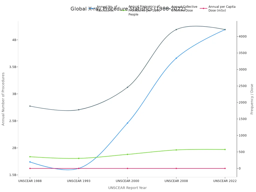
Artificial intelligence revolutionizes the 125-year-old X-ray. It transforms this foundational medical test into a powerful tool for proactive disease detection. The sheer volume of annual procedures highlights the technology's global impact.
Advanced algorithms analyze scans for subtle patterns invisible to the human eye. This capability is crucial, as perceptual errors account for 60-80% of interpretive mistakes. The modern ai x ray enables earlier diagnosis, improving patient outcomes.
How AI Enhances X-Ray Diagnostics

Artificial intelligence does not replace the expert radiologist. Instead, it serves as a powerful collaborator, augmenting human expertise to deliver faster, more accurate, and more comprehensive diagnoses. AI systems analyze medical images with a level of detail and consistency that complements the radiologist's critical judgment, transforming the diagnostic workflow from end to end.
A Digital Assistant for Radiologists
AI platforms function as a tireless digital assistant, streamlining workflows and enhancing diagnostic precision. These systems tackle repetitive tasks, reduce cognitive load, and help mitigate common human errors. By flagging subtle findings that might be missed during a busy shift, AI provides a crucial second look.
This collaboration yields significant improvements in accuracy.
- Physicians aided by an AI system show major gains in detecting chest X-ray abnormalities.
- Non-radiologist physicians using AI can detect abnormalities with the accuracy of a specialist, but in less time.
- In breast cancer screening, AI assistance decreases false positives by 37.3% and reduces unnecessary biopsies by nearly 28%.
AI also optimizes the radiologist's time. By pre-analyzing images, it can organize worklists to prioritize the most critical cases. For example, an algorithm can instantly identify a potential pneumothorax (collapsed lung) in an emergency room X-ray. It then flags the scan for immediate review, reducing treatment delays. Studies show this assistance dramatically cuts down reading times. One analysis of chest radiographs found that AI support reduced the mean reading time from 24 seconds to just 12 seconds. This efficiency allows radiologists to focus their attention where it is needed most.
Learning from Millions of Scans
The power of a modern ai x ray system comes from its training. Developers train deep learning models on millions of anonymized X-ray images from diverse patient populations. This immense dataset allows the AI to learn and recognize infinitesimal patterns, textures, and density variations that are often invisible to the human eye.
Protecting Patient Privacy Using vast medical datasets requires strict ethical and security measures. To protect patient privacy, developers use techniques like:
- De-identification: Removing all personal identifiers from the images and associated data.
- Federated Learning: Training AI models on data at different locations without centralizing it, minimizing the risk of a large-scale data breach.
- Differential Privacy: Adding mathematical "noise" to obscure individual data points while preserving overall statistical patterns.
These trained models can enhance image quality itself. AI algorithms excel at digital "cleanup" to produce a clearer picture for the radiologist.
- Denoising: Techniques using Convolutional Neural Networks (CNNs) can reduce image noise by up to 50%, clarifying anatomical structures.
- Artifact Reduction: AI removes visual distortions caused by patient movement or metallic implants, improving the contrast-to-noise ratio by around 30%.
- Super-Resolution: AI can enhance the resolution of low-dose X-rays, making them comparable in quality to full-dose images. This capability helps reduce a patient's radiation exposure without sacrificing diagnostic clarity.
Opportunistic Disease Detection
Perhaps one of the most revolutionary applications of AI in radiology is opportunistic screening. This is the practice of using a patient's existing medical image, ordered for one specific reason, to screen for other, unrelated conditions. A single ai x ray of the chest, for instance, might be taken to investigate a cough. An AI algorithm can simultaneously analyze that same scan for signs of osteoporosis, aortic calcification, or liver disease.
This approach transforms routine imaging into a powerful tool for population health. It finds diseases early in patients who are not actively seeking a diagnosis for that condition. The economic benefits are also compelling.
A German study analyzed the cost-effectiveness of using AI for opportunistic osteoporosis screening on chest radiographs. The results were remarkable.
| Metric | Result | Interpretation |
|---|---|---|
| Intervention | AI-driven osteoporosis screening | For women aged 50+ |
| ICER | €13,340 per QALY gained | Highly cost-effective |
| Threshold | €50,000–€60,000 per QALY | The intervention is well below the accepted value threshold |
This data shows that opportunistic screening is not just a clinical breakthrough but also an affordable and scalable solution. It helps address widespread underdiagnosis of conditions like osteoporosis, ultimately reducing the long-term healthcare costs associated with fractures and other complications.
Breakthroughs in AI X Ray Technology
The theoretical promise of AI in radiology is rapidly becoming a clinical reality. Recent breakthroughs are not just incremental improvements; they represent fundamental shifts in how machines perceive medical images. These advanced systems now detect diseases earlier, identify risk factors for future conditions, and learn with a sophistication that rivals, and in some cases exceeds, human experts.
Spotting Lung Cancer and Heart Disease
Early detection is paramount for improving survival rates in lung cancer and managing cardiovascular disease. AI is making significant strides in both areas by analyzing standard chest X-rays with unprecedented accuracy.
For lung cancer, AI models excel at finding tiny, suspicious nodules that the human eye might miss. When comparing performance, the data is compelling.
| Metric | AI Models Range | Radiologists Range |
|---|---|---|
| Sensitivity | 60.58% to 93.3% | 76.27% to 86.7% |
| Specificity | 64% to 95.93% | 61.67% to 84% |
AI models consistently show higher sensitivity, meaning they are better at correctly identifying nodules. One algorithm achieved 92.3% sensitivity, finding 14 nodules that at least one radiologist had missed. While this can lead to more false positives (lower specificity), this trade-off ensures fewer cancers go undetected. The technology enhances a radiologist's perception by:
- Improving Lesion Conspicuity: AI highlights faint pleural lines, hairline fractures, and barely perceptible lung nodules, reducing diagnostic uncertainty.
- Prioritizing Critical Cases: The system can flag scans showing signs of a collapsed lung (pneumothorax) or fluid buildup (pleural effusion), directing immediate attention where it is needed most.
- Enhancing Consistency: AI uses structured outputs like heatmaps and bounding boxes to standardize how findings are reported, reducing variability between different readers.
Beyond cancer, AI can assess cardiovascular risk from the same chest X-ray. Deep learning models analyze the image for signs of Coronary Artery Calcification (CAC), a key indicator of heart disease risk. This turns a routine ai x ray into an opportunistic screening tool. It identifies at-risk patients without requiring a dedicated, higher-radiation CT scan. This approach is cost-effective, exposes patients to less radiation, and allows health systems to reuse millions of existing images to stratify population risk.
Detecting Liver Disease from Chest X-Rays
The power of opportunistic screening extends to conditions seemingly unrelated to the chest. Researchers have successfully trained AI models to detect fatty liver disease from chest radiographs. This is a remarkable feat, as the liver is only partially visible at the bottom of a standard chest X-ray.
The results are impressive. These models demonstrate high diagnostic performance, achieving an accuracy (measured as AUC) between 0.82 and 0.83. How does the AI do it?
Saliency maps, which visualize where the AI is "looking," show that the model focuses its attention on the liver and diaphragm region in over 74% of cases. It learns to associate subtle texture and density patterns in that specific area with the presence of liver disease.
This capability opens a new frontier for public health. It allows for the screening of a widespread, often silent condition using an imaging test that millions of people already receive for other reasons, such as a cough or chest pain.
Building Foundation Models like Ark+
The latest evolution in medical AI is the move from single-task algorithms to powerful "foundation models." A foundation model is trained on massive, diverse datasets to acquire a broad, general understanding of medical images. This pre-trained knowledge can then be quickly adapted (or "fine-tuned") for a wide range of specific diagnostic tasks.
Several organizations are at the forefront of developing these powerful tools.
| Company | AI X-ray Technology/Product |
|---|---|
| VUNO | VUNO Med®-Chest X-Ray (thoracic disease detection) |
| Siemens Healthineers | AI-Rad Companion (assists in identifying abnormalities) |
| Zebra Medical Vision | AI-driven imaging platform with multiple FDA-approved tools |
| Philips Healthcare | Advanced Visualization Workspace (integrates imaging modalities) |
A groundbreaking example is Ark+, a foundation model developed at Stanford. Unlike previous models that often discarded expert input, Ark+ is built using a method called "fully supervised learning."
"We wanted AI to learn from expert knowledge, not only from the raw data," explains researcher Eric Liang. Ark+ was trained on numerous datasets containing detailed annotations from expert physicians. This approach allows the AI to "learn from the human art of medicine," building a deeper, more nuanced understanding of disease.
This new generation of models offers transformative capabilities:
- Flexibility: They can be fine-tuned to identify new or rare diseases with very few examples, without needing complete retraining.
- Fairness: Training on diverse global datasets helps reduce inherent biases and ensures the model performs well across different patient populations.
- Accessibility: The code for models like Ark+ is often shared openly, allowing researchers worldwide to adapt it for local needs, even in clinics without large data resources.
Foundation models represent the future of medical AI. They move beyond simple detection to enable true clinical intelligence, paving the way for a smarter, safer, and more accessible healthcare system.
The ai x ray is pivotal in shifting medicine from a reactive to a proactive model. It enables population-scale screening and personalized risk assessment. This powerful fusion of AI and radiology empowers doctors, heralding a future of earlier, more precise, and accessible healthcare for everyone.
FAQ
Will AI replace radiologists?
No. AI acts as a powerful assistant. It augments the radiologist's expertise, handling repetitive tasks and highlighting subtle findings to improve accuracy and efficiency.
Is patient data safe during AI training?
Yes. Developers use strict privacy measures like de-identification and federated learning. These techniques protect patient information while allowing the AI to learn from diverse medical data.
How does an AI x ray find diseases?
The AI system trains on millions of annotated scans. It learns to identify subtle patterns and density variations that correlate with specific diseases, often before they are human-visible.
See Also
Top Chinese Suppliers for Acquiring X-Ray Inspection Equipment
Unveiling the Truth: Canine Influenza Virus Antigen Test Accuracy
Evaluating Investment in Automated Virus Sampling Tube Assembly Lines
Understanding Pharmaceutical Checkweighers: Key Definitions and Core Features
Computer Chips: Insights into Designer Ingenuity and Creative Minds